The same number twice, and two different cells
Two frames twenty-four minutes apart hold the same amount of condensed chromatin, to within two pixels. One is a cell going into mitosis and the other is a cell coming out of it, and only the picture says which.
A human cell divides in about an hour. A figure of it is one frame.
This one was imaged every 90 seconds for 60 minutes by confocal microscopy, triggered automatically when the cell entered prophase, and the whole stack was deposited. So it is possible to ask what any single frame of it would have left out.
-
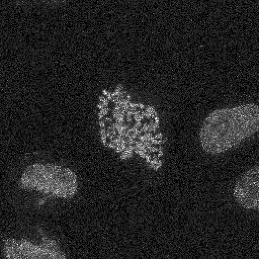
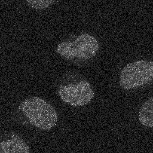
The cell has just entered prophase. The chromatin is spread through the nucleus and there is little that is bright.
Frame 0 of 40, 0 minutes in. 280 pixels above the cut.
-
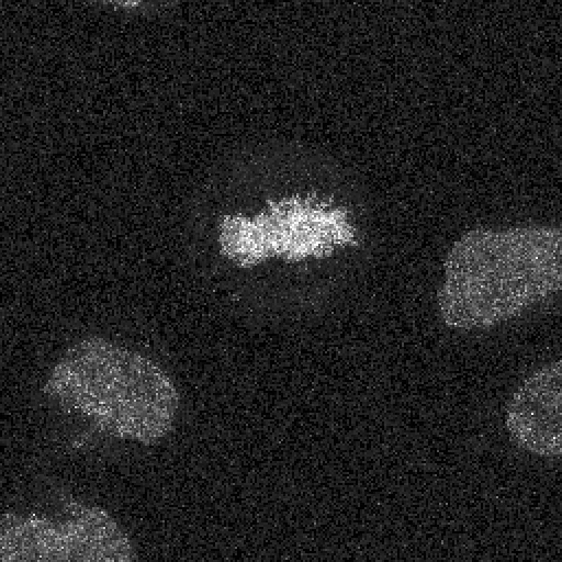
Fifteen minutes in. The chromosomes are condensing: the same DNA, packed into less space, so each pixel holding it is brighter.
Frame 10 of 40, 15 minutes in. 487 pixels above the cut.
-
Twenty-seven minutes. This is as condensed as it gets, and it is the frame a figure would almost certainly use.
Frame 18 of 40, 27 minutes in. 665 pixels above the cut.
-
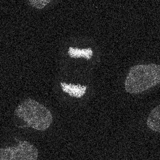
Forty-three minutes. Two separate masses, pulling apart. The amount of condensed chromatin is already falling.
Frame 29 of 40, 43.5 minutes in. 134 pixels above the cut.
-
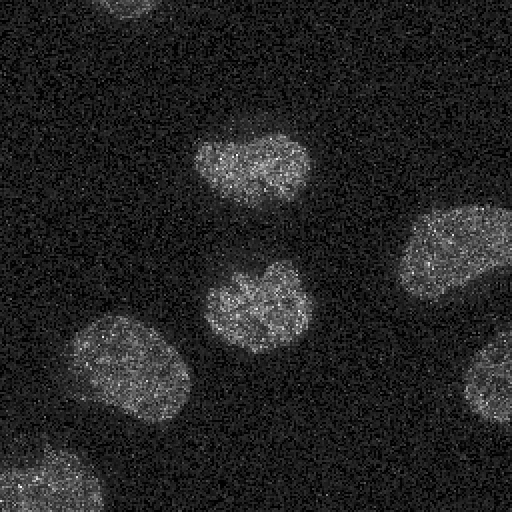
Fifty-eight minutes. Two daughter nuclei, decondensed, and almost nothing above the threshold any more.
Frame 39 of 40, 58.5 minutes in. 64 pixels above the cut.
One cell, one hour, five of forty frames
The DNA channel, projected over all 31 optical sections at each timepoint. Frames are taken at even positions through the series rather than chosen for what they show, so the sequence is sampled rather than curated. The other nuclei in the field belong to cells that are not dividing and sit still for the hour.
What rises and falls
Condensed chromatin is brighter per pixel than the same DNA spread through an interphase nucleus, so counting the pixels above a fixed intensity follows condensation without anything having to be outlined or identified.
It climbs to a peak at 27 minutes and falls away. That peak sits in the same place at every cut tried: 28.5 min, 27 min, 27 min at the 99.0th, 99.5th, 99.9th percentiles respectively. The height of the curve changes with the cut. Where the top of it falls does not.
The two frames this piece is about
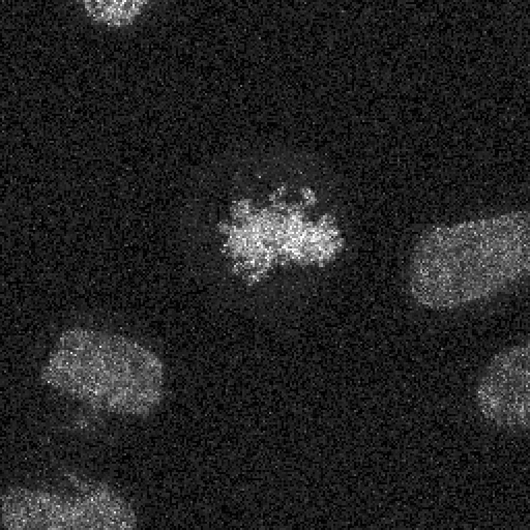
7.5 min, on the way in · 415 px
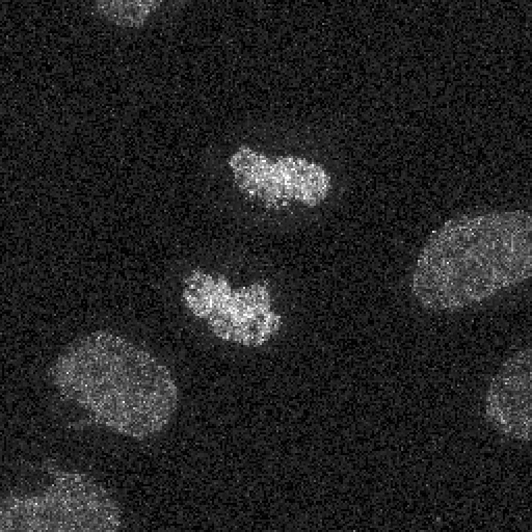
31.5 min, on the way out · 417 px
Both frames hold the same amount of condensed chromatin: 415 pixels above the cut on the left and 417 on the right, a difference of 2. They are 24 minutes apart and the cell is doing the opposite thing in each. The measurement cannot separate them. Nobody looking at them needs it to.
| Cut | Intensity | Peak | First frame | At the peak | Last frame |
|---|---|---|---|---|---|
| 99.0th percentile | 23369 | 28.5 min | 628 px | 990 px | 260 px |
| 99.5th percentile | 27380 | 27 min | 280 px | 665 px | 64 px |
| 99.9th percentile | 36190 | 27 min | 16 px | 255 px | 2 px |
A quantity that is the same in both. A cell that is doing opposite things in each. The number is not wrong, and it is not even imprecise: those two frames really do hold the same amount of condensed chromatin. It is simply not a quantity that distinguishes going in from coming out, and no amount of decimal places would make it one.
The pictures separate them in about a second, and they do it without anybody deciding in advance what to measure. That is the thing a visualisation is for which a table is not: it carries the properties nobody thought to ask about.
One caution about those two frames. The site measured a second quantity, the number of separate bright regions in each, and it comes out at 48 on the left and 23 on the right. That is a real difference and it is not a count of chromosomes or of chromosome masses: at these thresholds the chromatin resolves into many bright fragments rather than two tidy bodies. It is reported because it differs, not because it names anything.
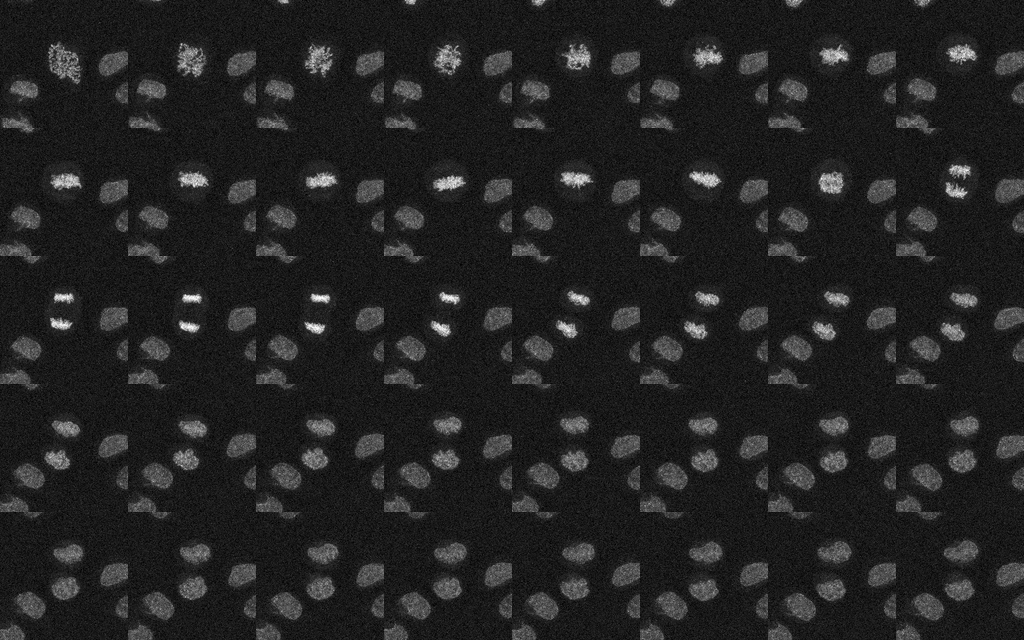
The whole series at once, in order, left to right and top to bottom. Each tile is one timepoint, ninety seconds apart. This is the figure that cannot be made from a single exposure, and it is the one that makes the direction obvious.
What this is not an argument for
Not against measuring. The curve above is a measurement, it is the thing that found the two frames, and no amount of looking at forty tiles would have turned up a pair matching to two pixels.
It is an argument against the order people usually put them in. The number is not the finding with the picture as decoration. Here the number raises the question and cannot answer it, and the picture answers it immediately. Both were in the same deposited file, free to anyone, for eight years.